The MINIONS (Patient-specific Microstructural and radIobiological model for persoNalised external beam radiatIONn therapy in localised tumorS) project is an advanced research initiative aimed at integrating patient-specific microstructural and radiobiological models into personalized radiotherapy. This approach focuses on a detailed characterization of the patient’s tumor, towards improved tumour control probabilities.
The primary objective of MINIONS is therefore to develop a patient-specific model that accurately represents the microscopic features of tumors and their interactions with radiation.
The project consists in a set of multidisciplinary activities which will be conducted in four inter-connected workpackages.
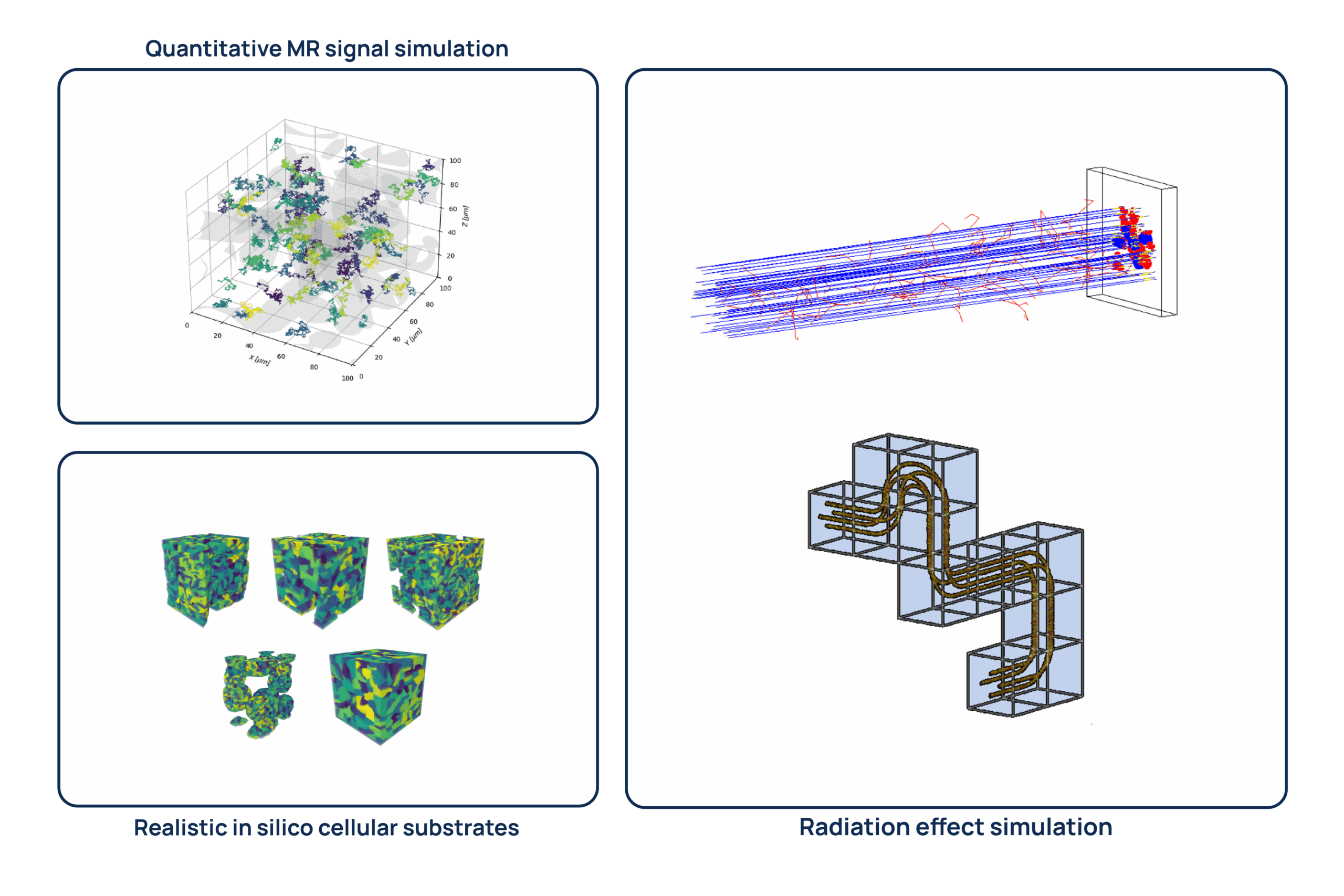
1. WP1 – Monte Carlo (MC) simulations of Quantitative Magnetic Resonance (qMRI) signals and radiation-tissue interactions over a library of realistic in silico cellular substrates to create an integrated simulation platform.
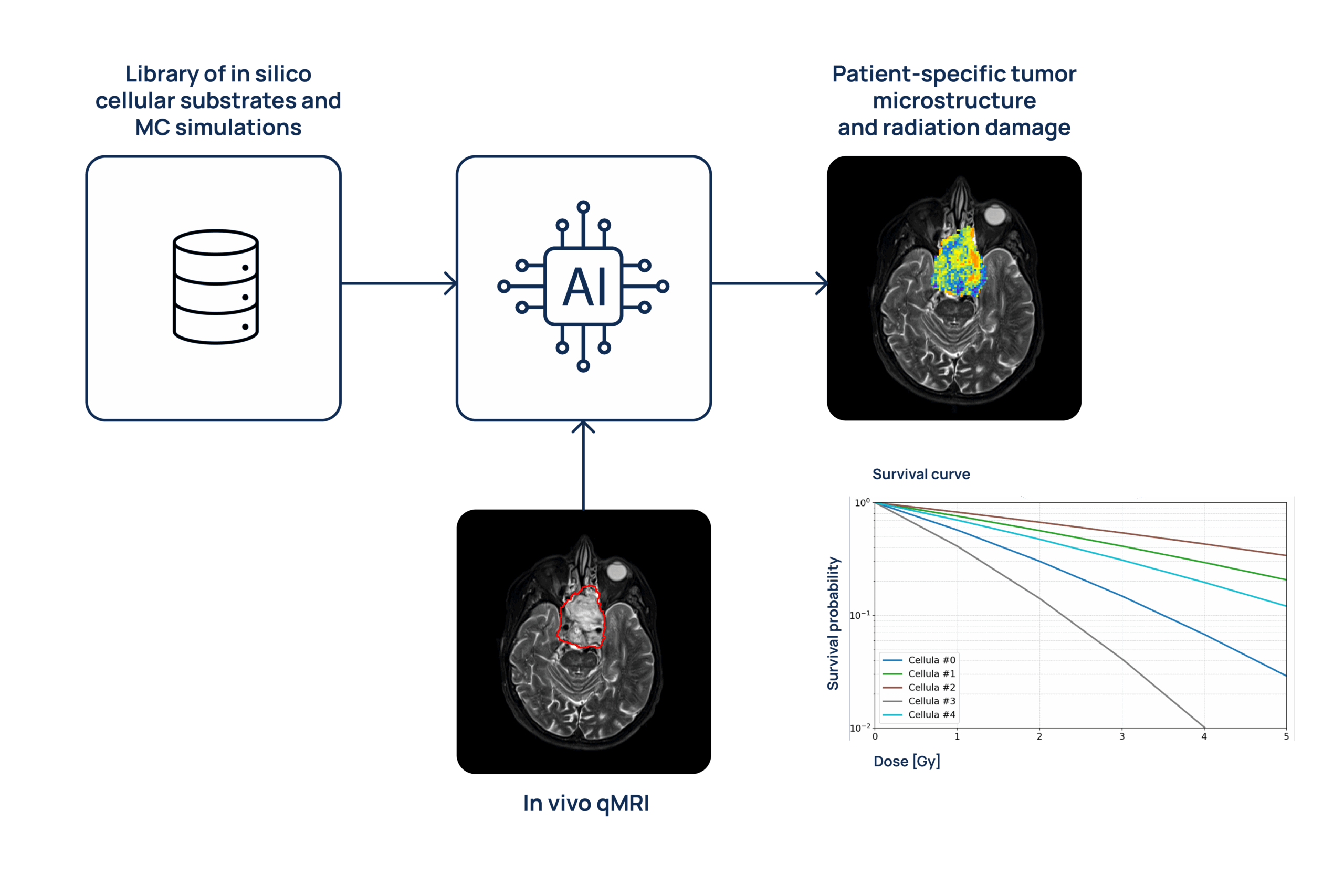
2. WP2 – Artificial Intelligence-based techniques to accelerate MC simulations, ensuring they are compatible with clinical workflows.
3. WP3 – Advanced qMRI imaging methods to non-invasively gather the necessary data to inform the simulation platform, enabling the development of patient-specific microstructural and radiobiological models.
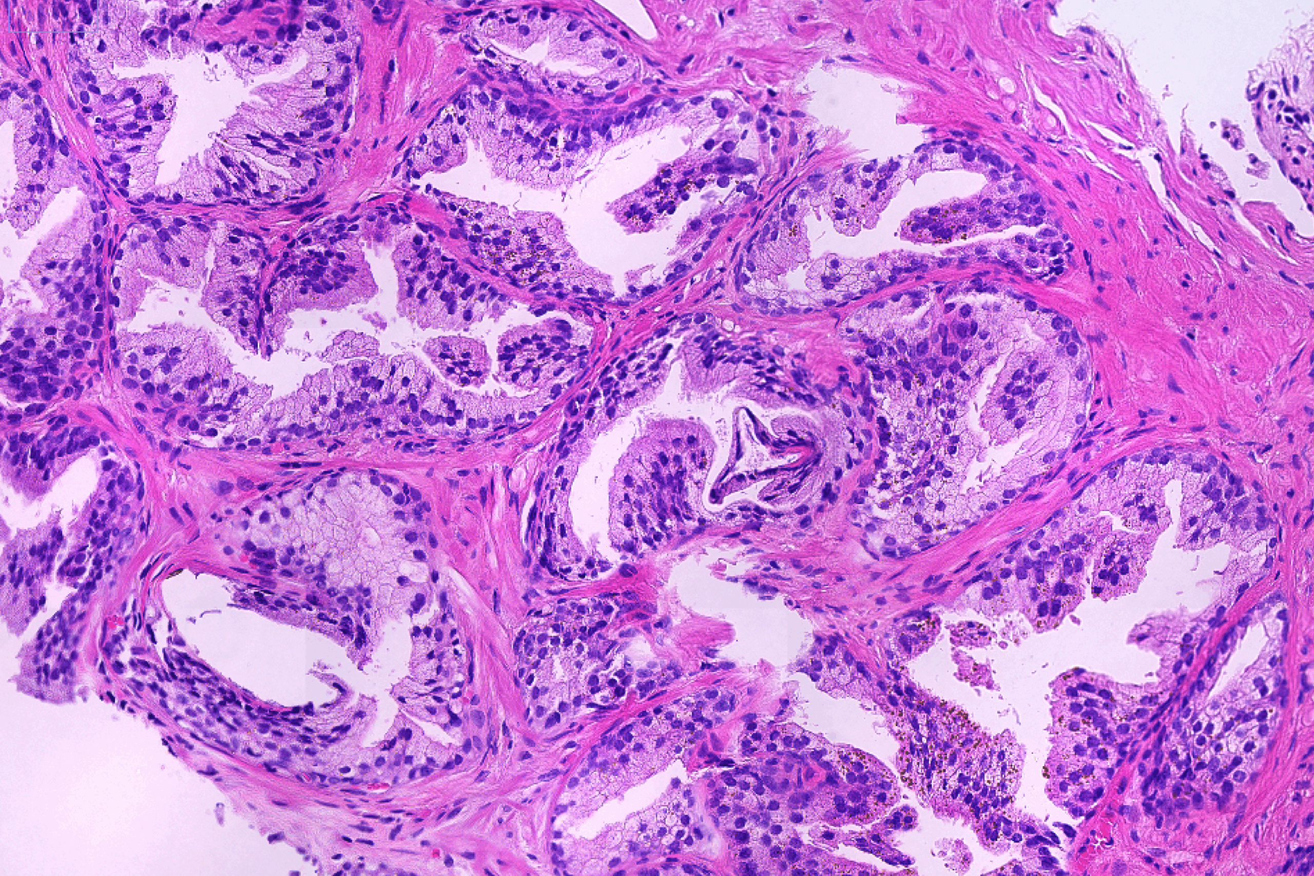
4. WP4 – Experimental validation relying on patient-specific histological samples as well as in vitro cells irradiation.
The expected result is the development of a patient-specific tumor model embedded in a scalable simulation platform, useful to advance knowledge and technology in bioengineering.
The project will also have an impact on other fields, such as medical physics, radiobiology, radiology and radiation oncology, towards the development of biologically targeted treatments.
The MINIONS project, funded by the European Union through an ERC Starting Grant (Chiara Paganelli, Principal Investigator), is carried out at the CartCasLab of the Politecnico di Milano.
WP1 – Development of an Integrated Monte Carlo (MC) Simulation Platform
Development of an integrated platform of realistic and dynamic in silico 3D cellular substrates coupled with simulations of qMRI signals and radiation interactions.
Tasks
Task 1.1 – Generation of Realistic In Silico Substrates
Starting from histological data, in vitro cell lines, and literature information, we will build a library of realistic 3D cellular substrates. These virtual tissues will be grown using Monte Carlo (MC) simulations to reproduce tumor development.
Dedicated simulations will model both morphological evolution, capturing realistic tissue architectures, as well as biochemical dynamics, such as oxygen diffusion and related microenvironmental effects.
Task 1.2 – Monte Carlo Simulations of Quantitative MRI (qMRI) Signals and Radiation Damage
Within the generated 3D substrates, two MC simulation environments will be developed and integrated:
- qMRI signal simulations, as for example modeling the diffusion of water molecules to simulate diffusion MRI and oxygen variations for susceptibility-weighted MRI.
- Radiation-tissue interaction simulations, including modeling dose deposition at the cellular level (microscale) and DNA damage processes at the nanoscale.
Task 1.3 – Creation of an Integrated In Silico Tumor Model Library
The simulations from Tasks 1.1 and 1.2 will be combined to produce a comprehensive library of in silico tumor models, linking structural, biochemical, and radiobiological properties.
WP2 – Implementation of a Clinically-Compatible AI Model
Acceleration of the simulation environment exploiting Artificial Intelligence (AI) solutions to derive clinically compatible microstructural and radiobiological models.
Tasks
Task 2.1 – Development of AI-Based Acceleration Models
Monte Carlo simulations are computationally demanding. To address this, AI-based models will be developed to:
- Accelerate the extraction of microstructural parameters from in vivo MRI data (see WP3), using neural networks trained on simulated data.
- Speed up MC simulations of radiation–matter interactions at both micro and nano scales, for both single-cell and multi-cellular systems.
Task 2.2 – Validation of AI Models on Simulated Data
The AI models developed in Task 2.1 will be tested on independent simulated datasets, taking advantage of the accurate ground-truth provided by MC simulations.
WP3 – Generation of Patient-Specific Microstructural and Radiobiological Models
Application of the developed microstructural and radiobiological models to in-vivo qMRI data for patient-specific tumor characterization and treatment outcome prediction.
Tasks
Task 3.1 – Retrospective Collection of Clinical Imaging Data
Imaging data from four tumor types will be collected at our collaborating clinical institutions to test our models on real-world in vivo data:
- Common tumors include prostate and breast cancers treated with conventional X-ray radiotherapy
- Rare tumors include chordoma and adenoid cystic carcinoma, typically treated with charged particle therapy.
Task 3.2 – Prospective Acquisition of Advanced Imaging Data
New, non-invasive quantitative MRI (qMRI) data will be acquired prospectively in collaboration with clinical partners. Advanced techniques, including diffusion imaging and susceptibility-weighted imaging, will be used to assess tumor microstructure and radioresistance.
Task 3.3 – Development and Evaluation of Patient-Specific Models
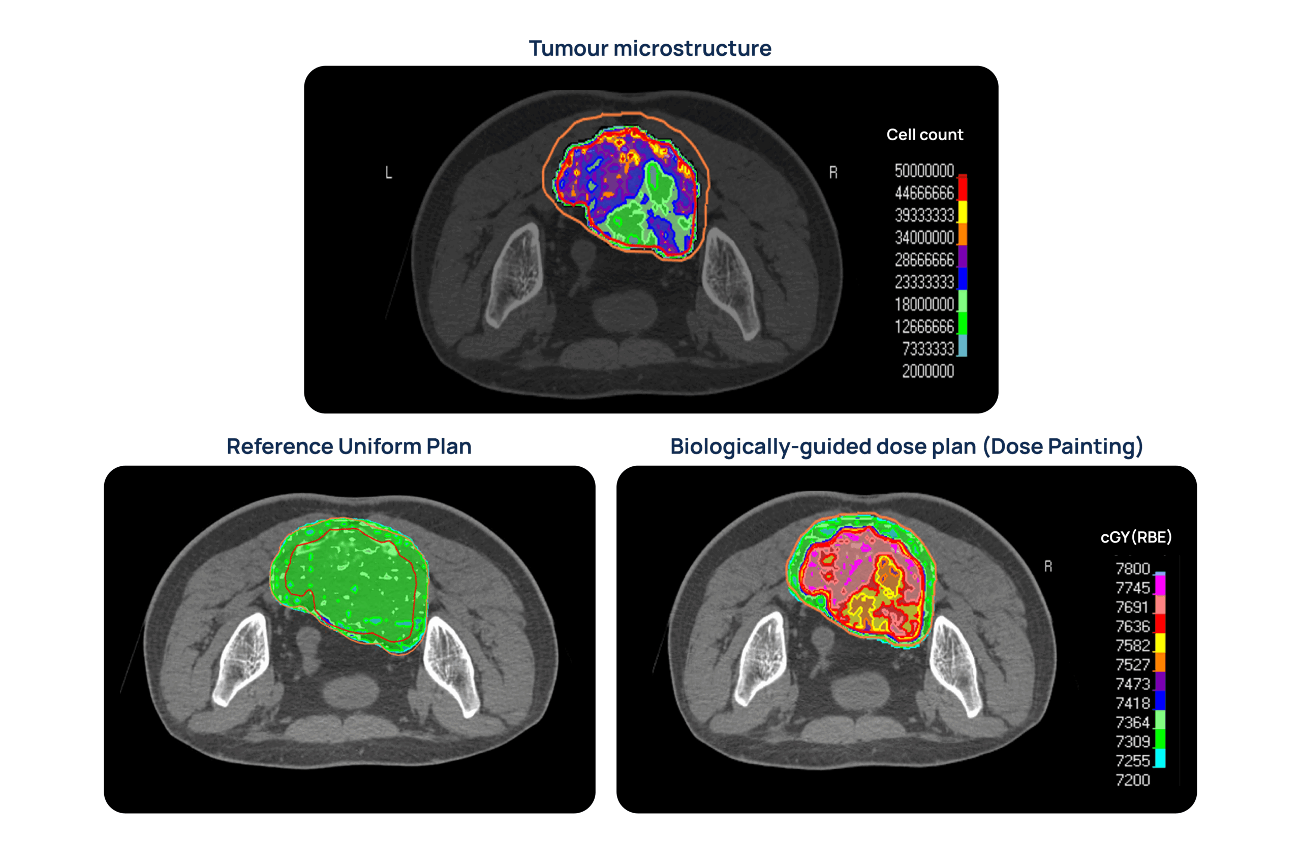
The AI and MC-based models trained in previous work packages will be applied to clinical datasets to derive personalized maps of tumor microstructure and radiobiology. Results will be compared with clinical information for correlation and predictive value. Moreover, the derived microstructural and radiobiological information will be used to implement personalized tumor control probability models towards biologically-guided dosimetric treatment plans.
WP4 – Validation of Patient-Specific Microstructural and Radiobiological Models
Validation of the microstructural and radiobiological models through histological analysis and in vitro irradiation.
Tasks
Task 4.1 – Validation with Histological Samples
During the prospective clinical protocol, biological samples will be collected and matched with in vivo MRI data, allowing direct validation of the microstructural features predicted by our models.
Task 4.2 – Experimental Validation with Irradiated Cell Cultures
From collected tumor samples, cell lines will be established and irradiated using X-ray and charged particle beams. This will enable direct validation of the simulated tissue-level and cellular-level radiation responses.